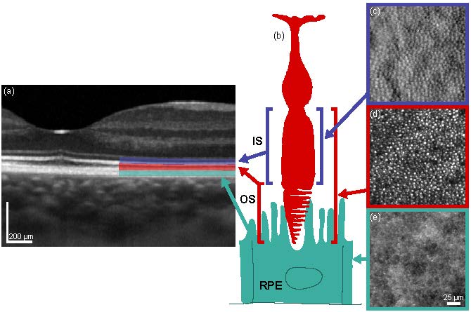
Adaptive optics imaging provides us with the ability to take microscopic pictures of the living human retina, revealing the structure of individual photoreceptors and retinal pigment epithelium (RPE) cells and the organization of their cell mosaics. We are combining live imaging of retinal cells with psychophysics to study the functional implications of normal and pathological structural changes in the eye’s photoreceptor and RPE layers, and how this relates to how we see colour, pattern and motion.

The current adaptive optics scanning light ophthalmoscope (AOSLO) was built in the spring of 2016. The system was designed for us by Alfredo Dubra and incorporates a split detector system. This gives us the opportunity to image and examine the wave-guided light reflex from the photoreceptor outer segments (cones and rods), the multiple scattered light from the cone inner segments and the retinal pigment epithelium cells in the dark-field images.
This instrument, a core facility of the research consortium , can image individual cone and rod photoreceptor cells (1–2 µm in size) and individual retinal pigment epithelial cells. It can also map the respective cell mosaics. It is non-invasive and allows monitoring in the authentic anatomical setting of the living human eye. This provides an unprecedented opportunity to prospectively study the earlies phases of AMD and to identify of new structural targets for the treatment of early AMD in human subjects that represent the transition between the anatomically pristine retina and early AMD.
A superluminescent diode (780 nm) forms a point source at the retina and the reflected light from this beacon is delivered to the custom Shack-Hartman wavefront sensor. The wave aberration of the eye is calculated, and a 97-degree-of-freedom deformable mirror (ALPAO DM97-15) changes shape to compensate for the eye's aberrations. High resolution retinal images are acquired after correcting for the eye's wave aberration. A superluminescent diode (840 nm) illuminates the retina, and an Exi-Aqua camera captures the light reflected from the retina.
Analysis of captured data is often performed through custom software, written in-house in C++ or Python, with user interfaces built in Qt. A variety of image processing techniques are used to semi-automatically identify photoreceptors and retinal pigment epithelium cells in AOSLO images, or retinal layers in OCT B-scans, to name two examples. Psychophysics stimuli use OpenGL for realtime rendering, on spectrally-calibrated monitors. Python, Octave and R Studio are also used for numerical analysis.